Specialities
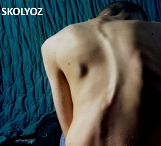
WHAT IS SCOLIOSIS?
Scoliosis is the general name given to the curvature of the spine to the right or left. When a healthy spine is viewed from behind, all vertebrae are aligned in the same direction. When this alignment is disrupted for any reason and an abnormal curvature occurs, it is called “scoliosis.” It would be more accurate to examine scoliosis under two separate categories: as pediatric-adolescent (or childhood-adolescent) scoliosis and adult scoliosis. Adult scoliosis can be a continuation of childhood scoliosis, or it can also occur as a result of wear and deterioration of the spine over time. Treatment options for adult and adolescent scoliosis are very different.
CAUSES OF CHILDHOOD AND ADOLESCENT SCOLIOSIS
Scoliosis can occur for a wide variety of reasons. For example, scoliosis can be seen in spastic children or in those who suffered a stroke during childhood. However, the most common types of scoliosis are idiopathic scoliosis — which occurs mostly in teens and whose cause is still unknown, and congenital scoliosis — which is caused by factors while the baby is still in the womb and is symptomatic from birth. The cause of idiopathic scoliosis is not known exactly. However, recent studies have revealed that some genetic factors play a role. Congenital scoliosis is thought to be caused by infections during pregnancy, diabetes and certain vitamin deficiencies.
The most common causes of scoliosis are as follows:
- Idiopathic (unknown cause) scoliosis. This occurs in a previously straight spine for an unknown reason. It is the most common cause of scoliosis.
Subclasses of idiopathic scoliosis: Idiopathic scoliosis is defined according to the age at which the scoliosis begins to develop. Each age group has its own characteristics and challenges in terms of treatment:
- Infantile-onset idiopathic scoliosis: 0-3 years
- Juvenile onset idiopathic scoliosis: 4-9 years
- Adolescent-onset idiopathic scoliosis: It is seen between the ages of 10-18.
- Congenital scoliosis: This is usually due to a defect in the spine formation during the intrauterine period.
- Neuromuscular scoliosis: This can be caused by conditions such as polio (polio), cerebral palsy or muscular dystrophy.
- Neurofibromatosis: This is a disease involving bone and soft tissues. Scoliosis is often associated with this condition.
- Rheumatic diseases
- Connective tissue diseases: Osteogenesis imperfecta, Ehlers Danlos, Marfan syndrome.
- Scoliosis may also develop after spinal fractures.
- Scoliosis after spinal infections
- Metabolic diseases: Diseases such as Morquio, Gaucher disease.
- Syndromic diseases: Scoliosis can also occur after many genetic syndromic diseases. Individuals with syndromic diseases should be closely monitored for scoliosis.
As is evident, there are many conditions that cause scoliosis. Therefore follow-up and treatment of patients can vary greatly according to the cause of the scoliosis and any underlying diseases.
WHAT ARE THE SYMPTOMS OF SCOLIOSIS?
Scoliosis is a disease that can threaten a child’s developing spine and put their lifelong spine health in jeopardy. When this disease is detected early, a high rate of success can be achieved with various treatment options. However, if spinal curvatures are not diagnosed or treated in time, they can progress over time and prevent normal development in children. In adulthood; lower back and back pain as well as heart and lung dysfunctions can be observed. Scoliosis can be recognized with careful evaluation. There are usually no complaints of pain in the early stages of scoliosis. Therefore, the diagnosis is often made by chance. The first finding is usually a visual disturbance. Poor posture, one shoulder being higher than the other, a prominent shoulder blade protrusion on one side, ill-fitting clothes (asymmetrical skirt or dress lines in girls) are often noticed by family members or teachers. One of the easiest ways to detect scoliosis is the forward bending examination where the child bends forward trying to touch the ground with both hands without bending their knees. While in this position, if there is visible rib protrusion or asymmetry in the hips or lower back, it could suggest possible scoliosis. Scoliosis is usually detected in patients who consult their doctor with these complaints.
WHAT IS APPLIED IN TREATMENT
The goal of treating spinal deformities is to provide a cosmetically correct, balanced and painless spine structure and to prevent additional problems that may occur over time. Early diagnosis ensures that the curvature is detected at a young age, when possible, and the necessary measures are taken to prevent progression. Each patient should be evaluated individually in both diagnosis and treatment. In other words, each patient’s treatment is unique. The course of scoliosis varies from patient to patient. Scoliosis may progress, remain at the same degree or improve. Anticipation of this process is one of the main factors determining treatment. In general, the onset of scoliosis at a young age, the degree of skeletal maturation, double curvatures, and a high angle of curvature may be warning signs that the patient’s scoliosis may progress. These patients are monitored more closely and receive more intensive treatment. Therefore, a child with scoliosis should go in for regular checkups to a specialist physician.
Although the treatment of scoliosis may vary depending on the type and extent of the scoliosis, the most commonly applied treatment methods are as follows:
- Observation: In patients with a curve below 20 degrees and whose skeletal development is nearing completion, observation and periodic checks alone are sufficient. Observation continues until the patient completes skeletal development.
- Schroth method: Corrective treatment based on the principles of the Schroth method is now practiced worldwide; this method essentially treats the deformity caused by scoliosis in three dimensions and includes specific exercise components for each component.
- Corset (Lumbar Brace) Treatment: The purpose of the corset (also known as lumbar brace) is to try to prevent the increase in the curvature. Corsets are especially effective in children with curvature above 25 degrees and who are still growing. For curvatures above 25 degrees, a corset treatment can be combined with Schroth exercises. The effect of the brace begins to decrease in curvatures over 40 degrees and in children whose skeletal development completion is still many years away.

- Surgical Treatment: If the patient’s degree of curvature is over 40-45 degrees and the child is still growing, surgery is inevitable. Curvatures over 50 degrees continue to increase even after growth has ended. For this reason, surgery is performed to prevent future health-related complications as well as for cosmetic correction. In some early-onset and congenital scoliosis, surgical treatment can be performed before the curvature reaches 50 degrees.

The “Vertebral Body Tethering (VBT) method” has also gained popularity in recent years and is starting to be used more commonly as a treatment option in scoliosis cases. Unlike spinal fusion surgery, this treatment method uses ropes and bands to prevent restriction of spinal mobility. Therefore, the child’s physical development is not hindered. On the contrary, as the child grows, this method allows for the remaining curvature to continue to correct itself. In other words, the main purpose of this method is to ensure that the spine is reshaped over time. With the help of ropes, the treatment is performed without restricting mobility. This way, new surgeries are not needed for the problems caused by the inhibition of spinal movements. Part of the curvature is initially corrected with surgery and the remaining degree of curvature is corrected as the development continues.
Who can benefit from VBT Surgery?
In order to undergo VBT surgery, the patient must meet the following criteria:
- Have a spinal curvature between 35 and 65 degrees
- Have a diagnosis of idiopathic scoliosis (meaning the scoliosis is not related to genetic or neurological diseases, injury or malformation)
- Be between 8-16 years of age
- Obtain confirmation by the doctor that bone development is not yet complete
- Have a need for surgical treatment for scoliosis
VBT surgery cannot be performed if the patient fulfills one or more of the following:
- Has complete bone development
- Has had previous spinal surgery
- Has a metabolic bone disease or unhealthy bones
- Has a spinal curvature of more than 65 degrees
- Has been diagnosed with kyphosis

SCOLIOSIS IN ADULTS
There are 2 main types of adulthood scoliosis:
- “Adult Idiopathic Scoliosis” which is scoliosis that started in childhood or adolescence and continues to progress during adulthood, and
- “Adult Degenerative Scoliosis” which is scoliosis that occurs with aging and deterioration of the spine over time.
Adult idiopathic scoliosis is painless in childhood but may cause pain in adulthood. Over time, patients themselves notice the imbalance and deformities in their torso. As back pain increases and movement capacity decreases, patients usually consult a doctor. Severe lower back and back pain may occur due to deterioration of the facet joints. Patients may have respiratory problems due to deformities in the thorax.
Adult degenerative scoliosis can be defined as scoliosis that occurs when the spine ages over time and deterioration ensues in the spine. It usually occurs over the age of 50. Over the age of 65 is the most common age period for adult degenerative scoliosis. Osteoporosis, i.e. bone resorption, can cause or contribute to scoliosis. Adult degenerative scoliosis is most common in the lumbar region, but can also occur in the neck and back. Due to deformities in the spine, patients notice shoulder and hip inequalities over time. As a result of these distortions, the patient’s torso may tilt sideways and forward. It has been shown that tilting the torso forward further impairs the patient’s comfort of life. The curvature of the spine causes the nerves coming out of the spine to be compressed in the canal, while thickening of the facet joints in the spine also contributes to nerve compression. This can lead to radiculopathy, or leg pain due to nerve compression, as well as muscle weakness and numbness. Over time, these disorders of the spine can cause one vertebra to slip forward (spondylolisthesis) or backward (retrolisthesis) in relation to the vertebra directly below it and can play a role as a contributing factor to the patient’s complaints of pain as well as nerve compression. X-rays, MRIs, computed tomography (CT) and electro-diagnostic tests (EMG) that show the entire spine can be used in the diagnosis of adult degenerative scoliosis.
The main factors to be considered when deciding on the treatment of adult scoliosis are the patient’s level of pain, the degree of the curvature and whether the curvature is progressing or not.
Non-surgical methods are preferred initially as a treatment option. While various physical therapy methods are applied under the guidance of a specialized physical therapist, painkillers may also be given for pain control. In addition, non-steroidal anti-inflammatory drugs (NSAIDs) can be added to the treatment to relieve irritation and inflammation, especially in facet joints or as a result of nerve compression. Transforaminal epidural steroid injections and facet joint injections are also good treatment alternatives for these patients. Each patient’s individual factors are taken into consideration when deciding on the sequence of these treatments.
Indications for surgical treatment in adult scoliosis are as follows:
- There is progressive scoliosis with or without pain
- Patient has persistent pain and significant loss of function despite non-surgical methods having been previously applied
- More immediate surgical treatment is especially considered in patients who have a narrow spinal canal with scoliosis and have difficulty in holding their urine and large bladder, which can be attributed to nerve compression.
Surgeries in adult scoliosis are more complicated than scoliosis in younger patients. There may be more procedures necessary and the duration of surgery may be longer. In elderly patients especially, accompanying problems such as heart-lung diseases, diabetes and osteoporosis may make procedures more difficult. However, very good results can be obtained when patients are well prepared for these surgeries under elective conditions.
Surgical treatment generally consists of removing the compression on the nerves (decompression), correcting the curvature enough to provide balance (instrumentation) and freezing the spine (fusion). The levels and extent of decompression and instrumentation are decided upon after a thorough evaluation of the patient’s complaints and after the areas under compression are determined and analyzed by MRI and computed tomography before surgery. In appropriate cases, the above-mentioned procedures can be performed with the help of small incisions with “Minimally Invasive Spine Surgery” methods.

KYPHOSIS
Kyphosis (hunchback) refers to an excessive curvature of the back. An inclination of 50 degrees or more at the level of the dorsal vertebrae is considered abnormal. When these degrees are exceeded, kyphosis deformity is present. When the patient is viewed from the side, his/her head is tilted forward and his/her back is bulging. Patients suffering from kyphosis usually complain of back and neck pain.
CAUSES
- Postural Kyphosis: (Kyphosis due to posture disorder) Postural kyphosis is usually detected in children during school screenings or by the family. It is more common in adolescent girls. The curvature is below 60 degrees and the spine is flexible. X-rays show no structural defect in the spine and the patient can correct the kyphosis at will. This condition usually improves with postural exercises and by changing postural habits.
- Scheuermann’s Disease: Scheuermann’s Disease is a structural hunchback of the spine at the back and back/waist junction that occurs in 0.5-9% of the population. It is more common in men. There are many theories about its causes, but the most widely accepted theory is that it is caused by malnutrition in the apophysis of the spine. It usually occurs during adolescence when growth is at its fastest. Patients consult a physician because of back or lower back pain or poor posture.

There are certain radiologic criteria for the diagnosis of Scheuermann’s Disease. Adolescent patients with mild kyphosis that does not progress below 50 degrees can be observed with X-rays taken every 4-6 months until growth is complete. In the conservative treatment of adolescents with a curvature between 50-80 degrees, the patient can first try to use a corset, but it is not common practice. Surgical treatment is considered if the curvature exceeds 80 degrees. In surgical treatment, only posterior instrumentation and fusion from the back of the spine is usually sufficient, but in very advanced and severe deformities, anterior release may be combined with posterior instrumentation and fusion. In such severe cases, we perform thoracoscopic anterior release (i.e. through 4 mini incisions with a camera system). Postoperative bracing is not necessary and the patient is ambulated as soon as possible. - Congenital kyphosis (congenital kyphosis): Congenital kyphosis develops due to congenital spinal abnormalities. The cause of the spinal abnormality may be a lack of formation, a lack of separation of the vertebrae or both. A “wedge spine” that develops anteriorly in the vertebral body due to lack of formation or a “half spine” (hemivertebra) that develops when the vertebral body is not fully formed anteriorly can cause kyphosis. Similarly, kyphosis can occur when the vertebrae are adjacent due to a lack of separation or when there is a band connecting the vertebrae also known as an unsegmented bar. Cases of congenital kyphosis due to lack of formation progress more rapidly and require early intervention than other types of kyphosis.
- Paralytic kyphosis: This kyphosis develops after neuromuscular diseases such as Polio disease and Cerebral Palsy.
- Myelomeningocele Meningocele and myelomeningocele, which develop after a closure defect of the spine, may result in the occurrence of kyphosis.
- Post-traumatic kyphosis: After spinal fractures, kyphosis may develop due to collapse and wedging of the anterior aspect of the vertebral body.
- Infectious/inflammatory causes: Infections such as tuberculosis and brucella are the most common infections that could cause kyphosis. Among inflammatory diseases, ankylosing spondylitis and rheumatoid arthritis are the most common diseases involving the spine.
- After surgical interventions: Kyphosis may develop after inadequate reduction or due to inadequate fixation during surgery.
- Metabolic or developmental causes: Kyphosis caused by diseases such as osteoporosis, osteomalacia and osteogenesis imperfecta, achondroplasia and mucopolysaccharidoses.
